For Appointments : 9867472415, 7715098366, 02249715624

Nuface Cleft and Maxillofacial Surgery

7715098366

  • Home
  • About Us
    • Dr. Parit Ladani
    • Dr. Jugal Toprani
  • TREATMENT
    • Cleft Lip and Palate
    • Corrective Jaw Surgery
    • Facial Asymmetry
    • Underbite Surgery
    • Genioplasty
    • V shape Jawline Surgery
    • Cyst and Tumour of Jaw
    • Wisdom Tooth Surgery
    • TMJ Disorders (TMD)
    • Rhinoplasty
    • Dental Implants
    • Ear Reconstruction
    • Obstructive Sleep Apnea
    • Buccal Fat Pad Removal
    • Face & Jaw Bone Fracture
    • Minor Oral Surgery
    • Facial Aesthetic Surgery
    • Double Chin Reduction
    • Dental Treatments
  • Gallery
    • Cleft Lip- Palate Surgery
    • Orthognathic Surgery
    • Cyst & Tumour of Jaw/Face
    • Unilateral Cleft Lip
    • Bilateral Cleft Lip
    • Why Cleft Lip occurs ?
    • Complication in Cleft ?
    • What is Cleft Palate ?
    • Speech Result
    • Youtube Videos
    • Jaw Surgery Cost In India
    • SURGERY VIDEOS
    • Lab Test in Cleft Patient
    • Cleft Lip-Dr Parit Ladani
    • What Is Cleft Palate ?
    • Cleft Lip Repair Surgery
    • Alveolar Bone Grafting
    • Symmetrical Nose
  • Contact Us
  • BLOG
  • More
    • Home
    • About Us
      • Dr. Parit Ladani
      • Dr. Jugal Toprani
    • TREATMENT
      • Cleft Lip and Palate
      • Corrective Jaw Surgery
      • Facial Asymmetry
      • Underbite Surgery
      • Genioplasty
      • V shape Jawline Surgery
      • Cyst and Tumour of Jaw
      • Wisdom Tooth Surgery
      • TMJ Disorders (TMD)
      • Rhinoplasty
      • Dental Implants
      • Ear Reconstruction
      • Obstructive Sleep Apnea
      • Buccal Fat Pad Removal
      • Face & Jaw Bone Fracture
      • Minor Oral Surgery
      • Facial Aesthetic Surgery
      • Double Chin Reduction
      • Dental Treatments
    • Gallery
      • Cleft Lip- Palate Surgery
      • Orthognathic Surgery
      • Cyst & Tumour of Jaw/Face
      • Unilateral Cleft Lip
      • Bilateral Cleft Lip
      • Why Cleft Lip occurs ?
      • Complication in Cleft ?
      • What is Cleft Palate ?
      • Speech Result
      • Youtube Videos
      • Jaw Surgery Cost In India
      • SURGERY VIDEOS
      • Lab Test in Cleft Patient
      • Cleft Lip-Dr Parit Ladani
      • What Is Cleft Palate ?
      • Cleft Lip Repair Surgery
      • Alveolar Bone Grafting
      • Symmetrical Nose
    • Contact Us
    • BLOG
Nuface Cleft and Maxillofacial Surgery

7715098366

  • Home
  • About Us
    • Dr. Parit Ladani
    • Dr. Jugal Toprani
  • TREATMENT
    • Cleft Lip and Palate
    • Corrective Jaw Surgery
    • Facial Asymmetry
    • Underbite Surgery
    • Genioplasty
    • V shape Jawline Surgery
    • Cyst and Tumour of Jaw
    • Wisdom Tooth Surgery
    • TMJ Disorders (TMD)
    • Rhinoplasty
    • Dental Implants
    • Ear Reconstruction
    • Obstructive Sleep Apnea
    • Buccal Fat Pad Removal
    • Face & Jaw Bone Fracture
    • Minor Oral Surgery
    • Facial Aesthetic Surgery
    • Double Chin Reduction
    • Dental Treatments
  • Gallery
    • Cleft Lip- Palate Surgery
    • Orthognathic Surgery
    • Cyst & Tumour of Jaw/Face
    • Unilateral Cleft Lip
    • Bilateral Cleft Lip
    • Why Cleft Lip occurs ?
    • Complication in Cleft ?
    • What is Cleft Palate ?
    • Speech Result
    • Youtube Videos
    • Jaw Surgery Cost In India
    • SURGERY VIDEOS
    • Lab Test in Cleft Patient
    • Cleft Lip-Dr Parit Ladani
    • What Is Cleft Palate ?
    • Cleft Lip Repair Surgery
    • Alveolar Bone Grafting
    • Symmetrical Nose
  • Contact Us
  • BLOG

Ear Reconstruction Surgery

Ear Reconstruction surgery using rib cartilage for microtia correction

What is Ear Reconstruction Surgery?

Ear reconstruction surgery is a surgery that creates a new ear or restores normal appearance to an injured or abnormally shaped ear. Ear reconstruction encompasses all operations used in the treatment of ear deformities. These conditions include birth defect of ear or missing ear (microtia), constricted ear deformities, protruding ears and traumatic (acquired) ear anomalies.  


A completely new ear can be made for someone who was born with an underdeveloped ear (microtia) or for someone who has had a severe injury to the outer ear. This may also be referred to as a total ear reconstruction. Other types of ear reconstruction may alter the existing cartilage and skin to restore normal appearance for prominent ears, constricted ears , Stahl ear deformity, cryptotia and ear lobe defect.

Why do you need ear reconstruction Surgery?

The auricle is a vital distinguishing feature of face, and hence its deformity has a profound effect on  appearance, self-confidence and psychological development in the afflicted children. Children born with microtia or other ear defects can lead full lives, especially with appropriate treatment and any needed lifestyle modifications. Talk to your ear reconstruction surgeon about the best course of action for you or your child.

Ear Reconstruction Surgery Involves correction of following ear deformity.

  • Microtia
  • Ear Lobe defect
  • Prominent Ear/ Bat Ear
  • Constricted Ear/ Cup Ear/ Lop Ear
  • Cryptotia 

Microtia Surgery

Normal structure of ear which helps in ear reconstruction for microtia

What is Microtia?

  • Microtia is a congenital malformation of the external ear when a baby is born with one or both ears that are significantly smaller than normal. The ear is usually so abnormally shaped and small that it no longer looks like an ear. Occasionally the ear may be completely absent, called anotia. 
  • When microtia is present the ear canal and ear drum are usually absent, a condition called aural atresia. 

GALLERY
types of microtia

Types of Microtia

There are several grading systems used to describe how severe the microtia is.

Microtia is classified into 4 grades:

  • Grade 1: The ear is slightly smaller than normal but retains most of the anatomic characteristics of the normal ear. Although atresia can be present, there is usually an external auditory canal.
  • Grade 2: The ear is even smaller and is less developed than grade 1. Part of the helix may be developed. The triangular fossa, scaphae, and antihelix are less developed. Atresia may or may not be present.
  • Grade 3 or classic: This grade is the most common type of microtia. It is usually a vertical remnant of skin consisting of a superior part that is made up of disorganized cartilage and an inferior part that is the displaced earlobe. Atresia is almost always associated with this type of microtia.
  • Grade 4 or anotia: There is complete absence of an outer ear structure. Atresia is almost always associated with anotia.

A simplified approach classifies the microtia as

  • Lobular - presence of ear remnant and lobule
  • Conchal - concha, external canal, and tragus present with lobule

Lobule type and Conchal type of microtia ear reconstruction

What Causes microtia?

  • Microtia usually occurs if there in an abnormality in the embryologic development of the six auricular hillocks. These develop at 4–12 weeks of gestation. The exact cause behind the abnormal embryologic development remains unknown.  
  • It appears to be more frequent at higher altitudes, especially above 2000 m, due to the low oxygen levels. 
  • However, certain teratogens like isotretinoin and thalidomide have also been implicated in this. 
  • Microtia is sometimes been linked to alcohol use during pregnancy, genetic conditions or changes, environmental triggers, and a diet low in carbohydrates and folic acid.

Ear reconstruction with Rib cartilage for lobule type microtia

How common is microtia?

The incidence of microtia is around 1 in 8000 - 10,000 live births. The condition is 2.5 times more common in boys than in girls and more commonly affects the right ear. Unilateral cases are four times as common as bilateral ones. Aural atresia is found with microtia in 75% of cases. 

Ear Reconstruction in Hemifacial Mircosomia

Can Mircotia be associated with other conditions or syndromes?

  • Microtia may be associated with other malformations such as hemifacial macrosomia, facial nerve weakness, urogenital defects, and cardiac and spine defects. 
  • It has also been associated with syndromes like Goldenhar syndrome and Treacher-Collins syndrome.

What to do if your baby is born without Ear?

best age for ear reconstruction

What to do if your baby is born with microtia?

If a child is born with microtia, then this should be followed by an early evaluation of the ear’s organ systems and the child’s hearing. The first consultation with ear reconstruction surgeon should be shortly after birth. This gives parents proper insight into their child’s condition and the treatment prospects. The management of a child with microtia should ideally be discussed with parents shortly after birth, to reduce parenteral stress and offer them reassurance. The microtia surgeon can discuss about all the possible option with the family, including observation or ear reconstruction with autologous costal cartilage, alloplasts, and prosthetics. Each method of management has a different timing, and therefore, all options must be discussed at an early stage. In addition, the microtia surgeon must also coordinate ear reconstruction with auditory management, so that the timing of the hearing correction may be optimized.

Hearing management in Mircotia children.

Unilateral Microtia

In unilateral cases, children generally retain full hearing in the unaffected ear. Sound can be absorbed into the still-functioning inner ear too. An audiogram test should be repeated at yearly intervals. A radiologist will evaluate the middle ear structures and ear canal using a CT scan. Treatment can be thoughtfully planned after consulting with relevant specialists.

Bilateral Microtia

In bilateral cases, children are at risk of greater hearing loss. This can lead to poor speech development. Children with bilateral microtia require earlier intervention. An audiogram is recommended within the first few days of life. A CT scan will evaluate whether the middle ear structures can be surgically reconstructed or not. Children with bilateral microtia often use bone-conducting hearing aids before a surgery.

Timing of hearing correction surgery

Ideally it should be done after microtia reconstruction, otherwise, the scar it produces may jeopardize the result of ear reconstruction.

What is the best age for ear reconstruction surgery?

The timing of ear reconstruction surgery will depend upon multiple factors such as the surgeon preference, method of ear reconstruction, physical growth, psychological and social consideration. 

The ears reach much of their mature size by age 7. Generally, by the age of 6, cartilage is sufficiently developed to provide an optimal primary framework. However, the older the child, the more cartilage is available for reconstruction. We recommend ear reconstruction after 9 years of age, when the chest measures at least 60 cm at the xiphisternum. 

Can Ear be Reconstructed?

Can Ear be Replaced or Reconstructed or Transplanted?

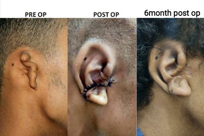
There are three main approaches to reconstruction of the external ear for treatment of microtia, each is discussed in detail on their respective sections. The two most popular methods involve burying a framework under the skin to create a new ear which will be a permanent part of the patient. 

The first, which is the oldest type of reconstruction, uses rib cartilage harvested from the chest that is carved and assembled into a framework with a shape similar to a normal appearing ear. Autologous rib cartilage transplantation is the current gold-stand treatment for microtia, but harvesting rib cartilage inevitably results in donor site injury. 

The second method uses an implant made from Porous Polyethylene (Medpor) to create a normal appearing ear however it lacks bioactivity and may cause extrusion and infection.  

The third method which is not part of the patient, is to use a removable or fix prosthetic ear that is attached to implants in the bone or applied to the skin with an adhesive.

Before the surgery, the surgeon will conduct a thorough history and examination, as well as hearing tests. The surgeon will also assess whether to use the person’s own (autologous) tissue, such as skin or cartilage or Medpor implants to reconstruct the ear or if a or prosthetic is more likely to yield a better result.

How long is ear reconstruction surgery take?

Ear reconstruction surgery is considered one of the most challenging facial reconstructive surgery procedures. The ear reconstruction process happens is in two stages, about 6-12 months apart. The first stage of ear surgery takes 4-6 hours and the expected length of stay in hospital is 5 days. The second stage takes 3-4 hours and the expected hospital stay is 2-3 days.

What involves in Ear Reconstruction Surgery?

Rib Cartilage for Ear Reconstruction Surgery

Rib Cartilage for Ear Reconstruction

How to Reconstruct Ear with Rib Cartilage?

  • The current gold standard for ear reconstruction is autogenous reconstruction because of its overall low complication rate. 
  • It involves a highly skilled and technically challenging task of harvesting the cartilage from the ribs and then carving out the framework of the ear from those pieces of rib cartilage. 
  • Several techniques have been adopted for autologous ear reconstruction, i.e. the Brent method and the Nagata method. We have simplified the technique by combining both Nagata and Brent technique for better results. 

GALLERY
Planning for ear reconstruction

Advantages of Rib Cartilage for Ear Reconstruction

  • While the autologous method of ear reconstruction requires more than one stage, one advantage is that using a patient’s own tissue decreases the risk of infection and eliminates the chance of rejection of foreign objects.
  • Very little long-term complications because it uses bodies own tissue
  • Withstands trauma very well
  • Generally no problem with contact sports
  • Excellent results are achieved in experienced hands

Harvesting Rib cartilage for Ear Reconstruction

Disadvantages of Rib Cartilage for Ear Reconstruction

  • Longer surgery
  • Two to three surgical stages may be required
  • Results are highly dependent on skill and experience of the reconstructive surgeon

First Stage: Harvesting rib cartilage

  • The surgical template is prepared from radiograph film using normal ear as a guide. In case of Bilateral Microtia, parents ear are used as a guide. 
  • Rib cartilage is harvested from the ipsilateral side through a small 5-cm long transverse incision on the skin over the rib cartilages. This gives the best possible cosmetic outcome for the patient and is specially useful in minimizing the scar in female children. 
  • The rib cartilage is harvested from 6th,7th,8th and 9th ribs. 
  • This part of surgery needs great patience and technical knowledge on the part of the surgeon, because it is very important to prevent future chest wall deformity. 
  • At the time of completion of operation a local anaesthetic nerve block is given to minimize the post-operative pain and discomfort.

Framework for Ear Reconstruction

Ear framework creation

  • During the initial stage, cartilage is taken from the ribs are used to create a three-dimensional sculpture that looks like a normal ear. 
  • Different portions of the cartilages are sculpted using precision instruments and joined together with the help of specially designed stainless steel wire sutures. 
  • If the patient has a normal second ear, it is used as a template to create the new ear from cartilage.

Framework placement in skin pocket for ear reconstruction

Pocket creation

  • A small incision is given at the site of designated ear position which should match with the normal ear on the other side. 
  • The deformed and redundant cartilage is excised carefully and with the help of specially designed scissors an ear pocket is created.

Ear Reconstruction using Rib Cartilage

Ear Framework placement

  • The cartilage framework is then placed under the skin at the site of the newly placed ear, to match the opposite ear. 
  • Additional cartilage may be saved either at the rib donor site or underneath the temporal scalp for use in a later stage. 
  • Suction catheters are placed under the skin to create better definition around the framework. Suction catheters are removed in 5 days. 
  • Children are usually admitted to the hospital for three to five days. 
  • At the completion of the first stage operation all the features visible in a normal human ear should be identifiable in the reconstructed ear.

Ear Reconstruction using Rib Cartilage

Post-operative care

  • Patient is nursed in a room and as the operative site settles, the skin stitches are removed by the 7th day after operation.
  • Children are allowed to walk on next day after surgery and given a normal diet according to their liking.
  • Most of the children are discharged on 5th day of operation.
  • Reconstructed site is kept clean by carefully washing with soap during the shower.
  • Chest scar is covered with antibiotic ointment ,it helps in improving the outcome of the scar with better cosmetic result.

Ear Reconstruction using Rib Cartilage

Precautions after first stage

  • Patient should not sleep on the side of surgery for three months
  • Use a soft pillow
  • Contact sports should be avoided for three months

Ear Reconstruction using Rib Cartilage

Second Stage

  • Additional stages of repair will follow after your child has fully healed and recovered from the first stage. This typically takes 6-12 months. 
  • In the next stage of reconstruction, the new ear framework is elevated off the side of the head to match the natural position of a normal ear. 
  • The piece of rib cartilage, which was put under the chest wall skin at the time of first stage operation is taken out, shaped and used as a support for the elevated ear framework by precisely suturing it behind the ear. It helps in maintaining the projection of the ear. 
  • This stage may require the use of a flap of tissue (fascia) from the area just below the scalp (this is scientifically known as temporoparietal fascial flap) and a skin graft to cover the raised framework. Skin grafts are usually taken from the opposite ear or from the scalp, and sometimes from the area above the collarbone or groin. The skin graft is sutured in place and dressing is applied. The dressing may need to be changed every alternate day for a week.

Ear Reconstruction using Rib Cartilage

Splint for Support

  • A special splint is provided to the patient at the end of second week. This helps in maintaining the elevation of the ear over long term and needs to be worn by the patient at all times other than bathing and cleaning for the duration of six to nine months.
  • If the patient wears spectacles and has difficulty in wearing the splint, the spectacle limbs could be modified to incorporate the splint thereby avoiding the problem.

Ear Reconstruction using Rib Cartilage

Precautions in the post-operative period

  • Patient should not sleep on the same side of operation
  • Contact sports are avoided for three months.


Excellent results could be achieved with this state of art technique if performed by an experienced surgeon.

Medpor Ear Reconstruction

Ear Reconstruction Using Medpor Implant

Total Ear Reconstruction with Medpor

An alternative technique for ear reconstruction uses a pre-fabricated artificial porous polyethylene (Medpor) framework to create the ear shape. This framework is then covered by a flap of tissue (fascia) from the side of the head that lies just below the scalp (temporo-parietal fascia flap) and a skin graft. Suction catheters may or may not be used postoperatively, and a large pressure dressing is placed. This procedure may be done as an overnight stay or occasionally on an outpatient basis.

Advantages

  • It is a single stage reconstruction
  • As patient’s cartilage is not required, surgery can be done at a much earlier age

Disadvantages

  • There is higher risk of implant exposure compared to autogenous cartilage reconstruction technique.
  • If the exposure occurs, it is very difficult to salvage the implant
  • The front portion of ear is covered by a skin graft, it does not give the normal kind of sensation to the ear as compared to the autogenous cartilage reconstruction
  • Medpor is a brittle substance and there is always a risk of fracture of implant and disfigurement in case of injury

Ear Reconstruction using Prosthesis

Prosthetic Ear Reconstruction

Who is a candidate for Prosthetic Ear Reconstruction?

Ear prosthetics, artificial ears attached by adhesives or bone implants, are another way to reconstruct ears. However, ear prosthetics are rarely suitable for children. The prostheses are delicate and expensive, and patient compliance and upkeep are very important to their use and success. They tend to be better suited for adults with acquired ear loss, as they lead a less active lifestyle than children, and are less likely to damage the prostheses.

Auricular prostheses are made for traumatic, surgical, and congenital defects. Surgical versus prosthetic reconstruction remains controversial for microtia patients. The ideal age for prosthetic treatment is between 6-9 years and is consistent with the optimal age for surgical reconstruction. The child should be mature enough to want and help care for the prosthesis. In cases of unsuccessful surgical outcome, the prosthetic ear is one of the option.  Prosthetic ear is a suitable option for the ear defects caused by trauma and disease in adult populations.

Retention of the prosthesis

Tissue adhesives are used conventionally for ear prosthesis retention because of their low cost and quick results. Tissue adhesive is an old method and causes skin reaction. It is difficult to wear and take off adhesive while bathing and swimming. 

Osseointegrated implant is superior option for retention and because of nature sense of prosthesis, ease of use, long life time and retention during daily activities is accepted by patients. An implant-retained prosthesis may also be advantageous when the defect has hair on the surface of where the prosthesis needs to be placed or when the patient has excessively oily and perspiring skin. A bar/clip mechanism or magnets are used as the interface of attaching the prosthesis to the implant screws. One contraindication to osseointegrated implants is radiation therapy treatment. After radiation therapy, the bone suffers from demineralization, hard tissue vasculitis, fibrosis, long-lasting infections, poor blood supply, and oxygen deficiency. Hyperbaric oxygen treatment allows irradiated patients to obtain osseointegration.

Firstly, Titanium implants are drilled in to the skull. Abutments are provided and on top of that prosthetic ears are fixed.

Indications

  • Only reserved for failed ear reconstructions with no other option remaining
  • Older people who are not fit for surgery

Problems

  • The ear has to be removed daily at the sleeping time, therefore, the patients are never able to incorporate the implant in there body image and a sense of deformity remains in the psyche
  • Implant sites need care by cleaning around regularly
  • Prosthetic ears lack sensations and the colour and temperature does not change with the body and patients are always able to perceive the difference
  • Needs to be changed every five years
  • The long term cost is higher than the autogenous cartilage reconstruction·  The ear has to be removed daily at the sleeping time, therefore, the patients are never able to incorporate the implant in there body image and a sense of deformity remains in the psyche
  • Implant sites need care by cleaning around regularly
  • Prosthetic ears lack sensations and the colour and temperature does not change with the body and patients are always able to perceive the difference
  • Needs to be changed every five years
  • The long term cost is higher than the autogenous cartilage reconstruction

How to find best Ear Reconstruction surgeon/Microtia surgeon

Choosing an Ear Reconstruction Surgeon for Microtia.

Ear reconstruction surgery is one of the most challenging operations to perform, due to the complex shape that must be created for the ear to look natural. The condition of microtia is not so common, and only a few doctors have the necessary knowledge, practice, and skills to treat the condition successfully. Only highly skilled & experienced Microtia surgeons can perform the surgery to get consistent results.


Dr. Parit Ladani completed his training under Dr. Z C Chen, world-renowned authority in ear reconstruction at Chang Gung Memorial Hospital, Taiwan. Dr. Parit Ladani is a well know surgeon for microtia and ear deformities in India. He is an expert in ear reconstruction surgery for children who are born with missing or partial ear or have had a traumatic loss of ear. 


He performs various ear surgeries, otoplasty, anotia, and deformed ear reconstruction. Dr. Parit Ladani is a highly skilled and experienced ear reconstruction surgeon and delivers his patients with consistent results. He has performed more than 100 ear reconstruction surgery till date.

Best Ear Reconstruction Surgeon

Dr. Parit Ladani preparing Ear Framework from Rib Cartilage

How much does Ear reconstruction surgery cost in India?

How much does ear reconstruction cost?

There are several factors that can affect the cost of microtia surgery. The extent and complexity of surgery can often dictate the final cost of treatment. The cost for ear reconstruction surgery depends on number of stages required for surgery, method of ear reconstruction (autologous rib cartilage reconstruction or MEDPOR reconstruction), Hospital or Surgical facility cost, Surgeon’s fee, Anaesthesia Fees, medicine cost and  investigations. A surgeon's fee for ear reconstruction surgery will be based on his experience, the type of procedure used and the geographic office location.


Cost of ear reconstruction surgery in India is only a fraction of what it costs in developed countries. The results are however as good as in any western country. Ear reconstruction surgery is offered at very affordable cost of 1.5-2 Lac for first stage and 75000-90000/- for second stage at Nuface Clinic.


When choosing a board certified oral and maxillofacial Surgeon for ear surgery, remember that the surgeon's experience and your comfort with him are just as important as the final cost of the surgery.

Cost for Ear reconstruction Surgery in India

Ear Reconstruction with Costal Cartilage

    Frequently asked questions

    Frequently Asked Questions on Ear Reconstruction

    1. What is the incidence of microtia?

    Microtia occurs in about 1 in 8000 to 10000 live births and is more common in males.

    2. What is the ideal age for microtia reconstruction?

    We recommend that 9-15 years of age as the ideal age to commence reconstruction. By this time the young patient’s cartilage is mature enough to withstand the shearing stress it is subjected to in a newly created skin pocket.

    3. How many stages would be required for completing the ear reconstruction?

    The number of stages would depend on the technique chosen for reconstruction after examining the patient. Generally a two-stage reconstruction protocol works well for most of the patients.

    4. Would there be a donor site chest wall deformity?

    On account of the our unique cartilage harvesting technique , there would be no chest wall deformity.

    5. What would be the total duration of treatment?

    The entire reconstructive regime would require about 9-12 months to conclude.

    6. Duration of hospital stay?

    First and second stage surgery both require about 3-5 days of hospital stay.

    7. When could patients resume their normal activities after surgery?

    After first stage - Normal activities could be resumed as soon as the chest and ear drains are removed, usually it takes around 3-5 days.

    After the second stage – It takes around a week for the skin graft to settle after the ear framework elevation, there after the patient could resume normal activities.

    8. Could this technique be also used for reconstructing ear loss following trauma or burns?

    Yes, this technique of ear reconstruction which is used for microtia could also be used for reconstructing ears after loss due to any other reason.

    9. Would I be able to get my ears pierced after surgery?

    Yes, piercing of the ear is possible after autogenous reconstruction of the ear.

    Otoplasty

    Otoplasty correction of Ear deformity

    What is otoplasty?

    Otoplasty is a type of cosmetic ear surgery that helps improve the shape, position or proportion of the ear. It is performed to correct defects of the ear that are present at birth but become apparent with development or to treat misshapen ears caused due to an injury. Hence, it is also known as ear reshaping or ear pinning. Examples are Prominent/Bat Ear, Constricted/cup/lop Ear, Cryptotia or Ear Lobe Defect.

    Who is a good candidate for otoplasty?

    Otoplasty is typically used for ears that:

    • protrude from the head
    • are larger or smaller than normal
    • have an abnormal shape due to injury, trauma, or a structural issue from birth

    Additionally, some people may have already had otoplasty and aren’t happy with the results. Because of this, they may choose to have another procedure.

    Good candidates for otoplasty include those who are having:

    • Ear Lobe defect
    • Prominent/Bat Ear
    • Constricted/Lop/Cup Ear
    • Cryptotia

    What is the best age for otoplasty?

    The ear is fully shaped at birth. By age three, the ear is almost 85% of the adult size. By age five or six, the adult size is nearly achieved. Further increase in size with aging is due to the elongation of the earlobe and not actual growth. Thus, the best age for otoplasty is after the adult ear size is achieved, which means after the age of five. Otoplasty procedure suitable after age of 5 yrs are, Prominent/bat ear surgery, Constricted/cup/lop ear surgery and Cryptotia correction. Ear Lobe defect can be repaired at any age.

    Is otoplasty permanent?

    This procedure usually creates permanent changes in your ears. If you follow our instructions after you get this procedure, your ears should maintain their new appearance for the rest of your life. Accordingly, this cosmetic technique is an excellent way to fix your ears in an efficient, cost-effective manner. Results from otoplasty are typically life-long, barring any incidents which impact the shape or proportion of the ears.

    What you can expect during Otoplasty?

    Before the procedure

    Otoplasty can be done in a hospital or an outpatient surgical facility.

    Sometimes the procedure is done with sedation and local anesthesia, which numbs only part of your body. In other cases, general anesthesia which renders you unconscious. 

    During the procedure

    Otoplasty techniques vary based on what kind of correction is needed. The specific technique your surgeon chooses will determine the location of the incisions and the resulting scars.


    Your doctor might make incisions:

    • On the backs of your ears
    • Within the inner creases of your ears


    After making incisions, your doctor might remove excess cartilage and skin. He or she will then fold the cartilage into the proper position and secure it with internal stitches. Additional stitches will be used to close the incisions.

    The procedure typically takes about two hours. 

    After the procedure

    After otoplasty, your ears will be covered in bandages for protection and support.

    You'll likely feel some discomfort and itching. Take pain medication as recommended by your doctor. If you take pain medication and your discomfort increases, contact your doctor immediately.

    To keep pressure off your ears, avoid sleeping on your side. Also try not to rub or place excessive force on the incisions. Consider wearing button-down shirts or shirts with loose fitting collars.

    A few days after otoplasty, your doctor will remove your bandages. Your ears will likely be swollen and red. You'll need to wear a loose headband that covers your ears at night for two to six weeks. This will help keep you from pulling your ears forward when rolling over in bed.

    Talk to your doctor about when — or if — your stitches will be removed. Some stitches dissolve on their own. Others must be removed in the doctor's office in the weeks after the procedure.

    Ask your doctor when it's OK to resume daily activities, such as bathing and physical activity.

    Are there any risk involved with otoplasty procedure?

    Otoplasty can lead to unwanted results:

    • Incomplete correction of prominent ears 
    • Telephone ear: This results if the mid-portion of the ear is corrected more than the upper and lower poles 
    • Reverse telephone ear deformity: This is a result of inadequate correction of the central portion of the ear relative to the upper and lower poles.
    • Overcorrection of a prominent ear can lead to abnormal appearance of the ear
    • Scarring
    • Swelling
    • Disfigured ears
    • Infection

    How to find best otoplasty surgeon in India?

    It’s important to choose your surgeon based on:

    • Education, training, and certification
    • Experience with ear surgery
    • Your comfort level with him or her

    Board certified Oral and Maxillofacial Surgeon are experienced and qualified to perform your aesthetic procedure. After finding a board-certified oral and maxillofacial surgeon in your area who is experienced in performing ear surgery, you will need to make an office appointment to set up your consultation. Dr. Parit Ladani is a board certified Oral and Maxillofacial Surgeon with experience of more than 15 year in field of ear reconstruction and otoplasty surgery. He has performed many complex ear reconstruction surgery. He is one of the best otoplasty surgeon in India.

    How much does otoplasty cost?

    It is imperative to note that there are various forms of ear deformities, hence the cost of treatment of each type of deformity is slightly different. At Nuface, we aim to correct the various types of ear deformities and offer a pleasing look for our patients. Dr. Parit Ladani is one of the best Oral and Maxillofacial Surgeon of India who is skilled in offering safe and aesthetically pleasing results. Dr. Parit Ladani has performed a large number of otoplasty surgeries with a very high success rate which has made him one of the best ear reconstruction surgeon in India. Nuface is one of the best Otoplasty clinic in Mumbai. To know more about the cost of Otoplasty, contact us or go to relevant section. (Ear Lobe Repair, Prominent/Bat Ear, Constricted/cup/lop Ear, Cryptotia)

    Ear Lobe Repair

    Can you repair torn earlobes?

    Ear lobe repair or auroplasty is a commonly performed out-patient procedure at Nuface Clinic. The hole in the delicate ear lobe tissue often gets stretched or may even tear through the ear lobule due to heavy ear rings or due to accidental traction injury. 

    GALLERY

    Why do the ear holes enlarge or split?

    Weight of the Earrings

    Heavy ear rings with Thin stalk or hook tend to cut through more easily. Thus an ear ring with a thicker hook or stud is preferable if your ear holes have a tendency to enlarge.


    Ageing

    During the natural process of ageing, the skin gradually loses collagen and elastin fibres which give strength, elasticity, and tightness to the skin.


    Trauma

    Sometimes earlobes may get cut because of trauma. Ear loops may be pulled by getting entangled in stuff, or maybe a small child pulls at it too hard.

    How do you fix a split earlobe?

    The procedure is performed under local anaesthesia. The skin edges of the enlarged hole or tear are shaved off to create raw edges. These edges are then meticulously sutured with fine threads to recreate an intact ear lobe. In some cases, additional manoeuvrers are done to recreate the round lower margin of the ear lobe. Closed the hole or tear along with leaving behind a small hole at the same time for wearing ear rings is not recommended as it leaves a weak spot in the resultant scar which is more likely to give way. A small dressing is applied.

    How long does ear lobe surgery take to heal?

    The dressing may be removed after 5 to 7 days. The sutures are removed after 7 to 10 days. The area heals very well and the scar is barely noticeable after a few weeks. 

    When Ear Piercing can be done after ear lobe repair?

    You must wait till the scar becomes strong enough before you pierce the ear lobe again. This duration may be as long as 3 to 6 months after surgery. Further, it is preferable that the new piercing is done away from the scar and not along the scar line.

    Which is better - Suture-less Glue repair vs Sutured Earlobe Repair?

    When correctly performed both types of repair can produce fantastic results. Though there are some caveats to either procedure.

    The suture-less glue repair may leave a scar that is not flush with the surface of the earlobe. The inwards dipping suture-line catches the light at the wrong angles and creates a shadow along the ridge making the scar very prominent. The incision scar may also widen with time.

    On the other hand, sutured repair causes the inconvenience of returning to the clinic for suture removal after seven days but produces a much better and thin scar which is generally flush with the surface of the earlobe.

    The reason is that sutures grab the skin as well as the soft tissue inside and hold them together while the tissues heal. Glue approximates the skin surfaces only, and the healing soft tissue beneath the skin shrinks away from each other causing valley like a deep final incision scar.

    How to prevent ear holes from enlarging?

    There is nothing you can do to change your skin type or eliminate gravity.

    Use lighter earrings for routine wear. Danglers (ornaments that hang) tend to pull on the ear lobe as they sway, so studs are a better choice as they stay snug to the ear lobe.

    Earrings with a thin central staff cut through the skin quicker than with thick stalk.

    You need to be more careful if you have thin skin and thin earlobes. Your earholes will probably enlarge early. But do remember that given enough weight all ear holes will expand eventually.

    How much does ear lobe repair surgery cost?

    Ear lobe repair surgery cost much lesser than western countries. Since it is out-patient procedure, cost for surgery is very much on the lesser side. Ear lobe repair surgery cost 5000/- to 10000/- per ear at Nuface Clinic.

    Ear Lobe Repair

      Prominent Ear/ Bat Ear Surgery In India

      Prominent Ear, Bat Ear Correction

      What is prominent Ear/ bat ear?

      Prominent ear also known as otapostasis or bat ear is an abnormally protruding human ear. In this condition, the concha (the depression in the external ear leading to its central opening) is large with poorly developed antihelix (a curved prominence of cartilage parallel with and in front of the helix on the pinna) and scapha (the curved depression separating the helix and antihelix of the ear). This results in the ear sitting further out from the head and is the most common problem causing prominent ears.


      Approximately 1-2 % of the total population is affected by prominent ears problem. It is an inherited problem and may be unilateral or bilateral and could be a result of lack or malformation of cartilage during primitive ear development. 

      GALLERY

      Is Prominent Ear bad?

      Having prominent ears may negatively affect a child’s self-image because he looks different and may be teased by peers. This can lead to poor development of interpersonal relationships, social withdrawal, and even depression.

      Is there any way to pin back ears without surgery?

      The ear cartilage in adults does not keep growing; it is fully mature and unfortunately, cannot be manipulated through any process other than surgery. The surgical correction however is a relatively easy procedure, low risk, quick recovery and very high reward.

      At what age can prominent ears be corrected?

      When planning surgery in children it is important not to interfere with growth so timing is important. Prominent ears should not be corrected surgically before this age, but can be successfully treated from 5 years onwards. By this stage the ear has achieved approximately 80% of adult size and the surgery will have little impact on future growth. 

      How to fix prominent Ear/Bat Ear?

      Surgery is required to treat prominent ears. This is usually performed under general anaesthetic, in hospital, as day surgery. It can be done as out-patient procedure under local anaesthesia for adults. Otoplasty involves exposing the cartilage structure of the ear and reshaping it to create the missing fold. Sutures are used to help hold the new shape.

      There are different types of surgical techniques to correct this defect, one used at our Centre is the Mustardè technique which is to perform a small incision behind the ear flap, a part of skin is removed so that you can access the cartilage which will be modified by weakening and if necessary removing the excess parts. The new shape is stabilized with the interior points and finally, the incision will be closed with stitches in absorbable material. Then the ear is immobilized in its new form with a bandage using an elastic band that will be kept for approximately 15 days. The surgery lasts for a total duration of about 60 minutes with a night of hospitalization and about two days of recovery.

      How much does it cost for Prominent/Bat Ear Correction Surgery?

      The cost of Prominent/Bat ear surgery varies from doctor to doctor and from one geographic area to another. Cost depends on surgeon fees, fees for the surgical facility, anesthesia, medical tests, prescriptions, surgical garments, or other miscellaneous costs related to ear surgery. Because otoplasty is elective surgery, insurance does not cover these costs. Approximate cost for Prominent/Bat Ear correction remains between 30000-100000/-.

      Choose your surgeon based on quality, training, and experience—not cost.

      Bat Ear / Prominent Correction

        Constricted Ear/Cup Ear/Lop Ear Surgery in India

        Constricted Ear, Lop Ear, Cup Ear Correction

        What are constricted/lop/cup ears?

        Constricted/lop/cup ears refer to a variety of ear deformities where the top rim of the ear (helical rim) is either folded over, wrinkled, or tight. This condition can range from mild to severe. In the mild form, the rim of the upper ear (helix) alone may be folded — this form is sometimes called lop ear. In the severe form, the cartilage of the helix and scapha (the curved depression below the rim of the upper ear) may be constricted, forming a tight roll.

        GALLERY

        What are the Causes of constricted/cup/lop ear?

        It is unknown what causes constricted ears. There may be a shortage of skin or cartilage, or both.

        How do fix constricted/cup/lop ear?

        Nonsurgical ear molding

        Newborns with constricted/lop/cup ear deformities may respond to non-operative molding while their ear cartilage is still soft. Ear molding uses a combination of a commercially available ear molding systems and orthodontic molding materials to reshape the ear.

        If ear molding is started early enough (ideally in the first few weeks of life), surgery can often be avoided. 

        Surgical treatment of constricted ears

        Once the ear cartilage has stiffened, surgery is necessary to correct the constricted ear deformity. Depending upon the severity of the condition, it may be possible to work with the ear cartilage already present to achieve a better shape of the ear. Constricted ear deformity, before and after surgery.

        In more severe cases of constricted/lop/cup ears, a rib cartilage graft may be necessary to replace wrinkled helical cartilage from the ear. If there is a tight band of skin that pulls the helix down, your child’s surgeon may add additional skin and cartilage from the normal ear to match the other side.

        Some children have bilateral constricted ears, in which both ears are affected. Your child’s surgical team will discuss options with you to determine whether to repair both ears at the same time, or in stages.

        How much does it cost for constricted/lop/cup ear correction surgery?

        The cost of constricted/lop/cup ear correction surgery varies from doctor to doctor and from one geographic area to another. Cost depends on surgeon fees, fees for the surgical facility, anesthesia, medical tests, prescriptions, surgical garments, or other miscellaneous costs related to ear surgery. Because otoplasty is elective surgery, insurance does not cover these costs. Approximate cost for Prominent/Bat Ear correction remains between 30000-100000/-.

        Choose your surgeon based on quality, training, and experience—not cost.

        Constricted Ear, Lop Ear, Cup Ear Surgery

          Cryptotia Ear Surgery in India

          Cryptotia Repair

          What is cryptotia?

          Cryptotia refers to an ear cartilage framework that is partially buried beneath the skin on the side of the head. The upper portion of the ear is hidden, and there may be some malformations of the cartilage of the upper ear (the scapha and the superior and inferior folds of cartilage, or crura). This can make it difficult to wear glasses or masks.

          What are the causes for cryptotia?

          It is unknown what causes cryptotia. It is more commonly seen among Asians. In Japan, it has been reported in as many as 1 in 500 births.

          How do you fix cryptotia?

          Non Surgical Treatment

          One treatment option for cryptotia is ear molding, a non-surgical treatment option that may help avoid the need for surgery. This treatment utilizes an external device that shapes and stretches the upper portion of the ear.

          Unlike many other congenital ear deformities, which are most effectively treated in the first few weeks of life when the ears are soft and mouldable, cryptotia may be effectively treated with ear molding well beyond the neonatal period. In some cases, successful non-operative correction of cryptotia has been reported even up to 5 years of age. This success may be possible because these ears already have a normal cartilage framework and primarily require skin expansion to cover the embedded cartilage.

          Surgical Treatment

          Cryptotia can also be treated surgically. Surgical correction of cryptotia may require the use of local skin flaps or skin grafts to lift the ear framework from the side of the head. Severe cases may require a cartilage graft.

          The goals of surgery are to:

          • Restore the auriculocephalic sulcus (the groove between the upper ear and the side of the head)
          • Replace the deficient skin using local skin flaps or skin grafts to cover embedded cartilage
          • Reinforce any collapsed cartilage
          • Release abnormal tethering ear muscles

          Although a general anesthetic is needed, the operation is done on an outpatient basis and your child will be able to return home the same day.

          How much does it cost for Cryptotia Ear Correction Surgery?

          The cost of Cryptotia surgery varies from doctor to doctor and from one geographic area to another. Cost depends on surgeon fees, fees for the surgical facility, anesthesia, medical tests, prescriptions, surgical garments, or other miscellaneous costs related to ear surgery. Because otoplasty is elective surgery, insurance does not cover these costs. Approximate cost for Prominent/Bat Ear correction remains between 30000-100000/-.

          Choose your surgeon based on quality, training, and experience—not cost.

          GALLERY

          Nuface Cleft and Maxillofacial Surgery Clinic

          B-105/106, Dhanashree Height, Bldg No. 42, Azad Nagar 2, Veera Desai Rd, Andheri W, Mumbai. 400053

          9867472415, 7715098366, 02249715624

          Copyright © 2022 Nuface Cleft & Maxillofacial Surgery Clinic - All Rights Reserved.

          Powered by

          This website uses cookies.

          We use cookies to analyze website traffic and optimize your website experience. By accepting our use of cookies, your data will be aggregated with all other user data.

          DeclineAccept